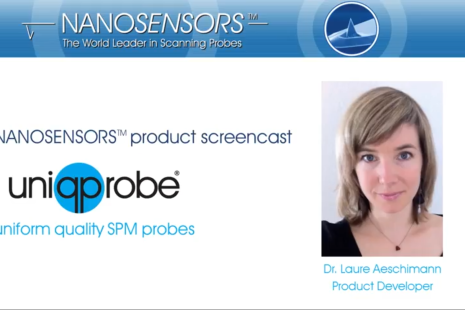
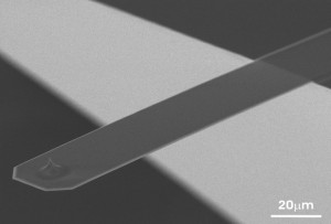
The screencast on the soft, drift-reduced NANOSENSORS™ uniqprobe cantilevers for biology and life science applications held by Dr. Laure Aeschimann has just passed the 1000 views mark. Congratulations Laure!
Since the first publication of this screencast that presents the uniqprobe types qp-BioAC, qp-BioT, qp-SCONT and qp-CONT , three further types of uniqprobe AFM probes have been introduced:
qp-BioAC-CI – a version of uniqprobe™ BioAC with Rounded Tips for Cell Imaging
qp-fast – three different uniqprobe™ cantilevers on one support chip for Soft- , Standard- , Fast Tapping/Dynamic AFM Imaging
and qp-HBC – the uniqprobe™ – HeartBeatCantilever that can also be used for ScanAsyst** and Peak Force Tapping** in Air.
To find out more please have a look at the NANOSENSORS™ uniqprobe brochure or the individual product pages on the NANOSENSORS webpage.
Additionally we have also put tipless versions of the qp-SCONT, qp-CONT and the qp-BioT ( SD-qp-BioT-TL, SD-qp-CONT-TL, SD-qp-SCONT-TL) and uniqprobe tipless cantilever arrays ( SD-qp-TL8a and SD-qp-TL8b ) on the NANOSENSORS special developments list.
Feel free to browse or let us know if you have any questions via info(at)nanosensors.com.
Product Screencast on the NANOSENSORS™ uniqprobe AFM Probes series with unsurpassed small variation in spring constant and resonance frequency by product developer Dr. Laure Aeschimann
A Japaneseversion of the screencast is also available :
Genetic and
environmental factors are key drivers regulating organismal lifespan but how
these impact healthspan is less well understood. Techniques capturing
biomechanical properties of tissues on a nano-scale level are providing new
insights into disease mechanisms.*
In the
article “ Mechanical properties measured by atomic force microscopy define
health biomarkers in ageing C. Elegans “ Clara L. Essmann, Daniel
Martinez-Martinez, Rosina Pryor, Kit-Yi Leung, Kalaivani Bala Krishnan,
Prudence Pokway Lui, Nicholas D.E. Greene, André E.X. Brown, Vijay M. Pawar,
Mandayam A. Srinivasan and Filipe Cabreiro apply Atomic Force Microscopy (AFM)
to quantitatively measure the change in biomechanical properties associated
with ageing Caenorhabditis elegans in addition to capturing high-resolution
topographical images of cuticle senescence.*
The authors
show that distinct dietary restriction regimes and genetic pathways that
increase lifespan lead to radically different healthspan outcomes. Hence, their
data support the view that prolonged lifespan does not always coincide with
extended healthspan. Importantly, they identify the insulin signalling pathway
in C. elegans and interventions altering bacterial physiology as increasing
both lifespan and healthspan.*
Overall,
AFM provides a highly sensitive technique to measure organismal biomechanical
fitness and delivers an approach to screen for health-improving conditions, an
essential step towards healthy ageing.*
The topographical images shown in this article were acquired using NANOSENSORS™ uniqprobeqp-CONT AFM probes. These AFM probes have very soft cantilevers designed for imaging of biological samples ( k = 0.1 N/m ).
Figure 1 f) from” Mechanical properties measured by atomic force microscopy define health biomarkersin ageing C. Elegans “ by Clara L. Essmann et al, please follow this external link for the full figure: https://www.nature.com/articles/s41467-020-14785-0/figures/1 f Representative AFM cuticle topography images of ageing C. elegans at different ages.
*Clara L. Essmann, Daniel Martinez-Martinez, Rosina Pryor, Kit-Yi Leung, Kalaivani Bala Krishnan, Prudence Pokway Lui, Nicholas D.E. Greene, André E.X. Brown, Vijay M. Pawar, Mandayam A. Srinivasan, Filipe Cabreiro Mechanical properties measured by atomic force microscopy define health biomarkers in ageing C. Elegans Nature Communications (2020) 11:1043 DOI: https://doi.org/10.1038/s41467-020-14785
Open Access: The article “Mechanical properties measured by atomic force microscopy define health biomarkersin ageing C. Elegans” by Clara L. Essmann, Daniel Martinez-Martinez, Rosina Pryor, Kit-Yi Leung, Kalaivani Bala Krishnan, Prudence Pokway Lui, Nicholas D.E. Greene, André E.X. Brown, Vijay M. Pawar, Mandayam A. Srinivasan, Filipe Cabreiro is licensed under a Creative Commons Attribution 4.0 International License, which permits use, sharing, adaptation, distribution and reproduction in any medium or format, as long as you give appropriate credit to the original author(s) and the source, provide a link to the Creative Commons license, and indicate if changes were made. The images or other third party material in this article are included in the article’s Creative Commons license, unless indicated otherwise in a credit line to the material. If material is not included in the article’s Creative Commons license and your intended use is not permitted by statutory regulation or exceeds the permitted use, you will need to obtain permission directly from the copyright holder. To view a copy of this license, visit http://creativecommons.org/licenses/by/4.0/.
High resolution imaging of the cytoplasmic side of bacteriorhodospin (BR) (left) and overlay of 6 force-distance curves (right) showing the controlled C-terminal unfolding of a single BR membrane protein from its native environment.
The measurements were recorded in buffer solution with a FlexAFM V3 (Nanosurf) using a NANOSENSORS™ uniqprobe qp-CONT cantilever by Dr. Patrick Frederix from Nanosurf AG.