Dynamics of Compartment-Specific Proteomic Landscapes of Hepatotoxic and Cholestatic Models of Liver Fibrosis
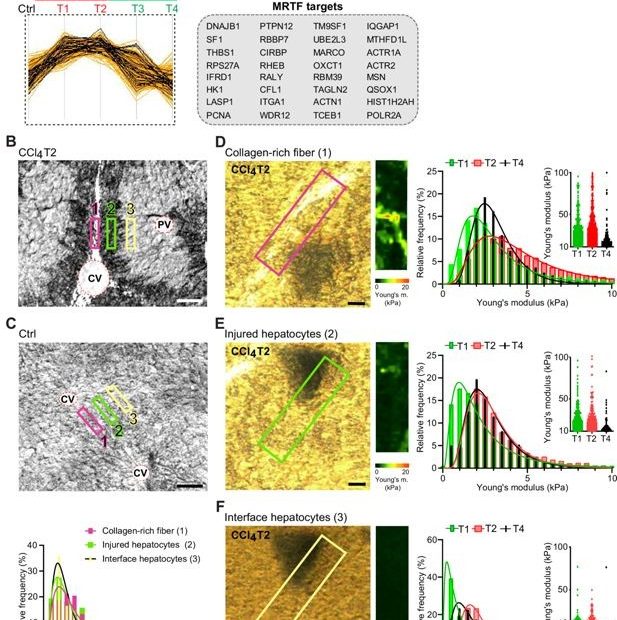
Liver fibrosis is characterized by excessive accumulation and remodeling of extracellular matrix (ECM) components, leading to altered tissue mechanics and progressive impairment of liver function.… Read More »Dynamics of Compartment-Specific Proteomic Landscapes of Hepatotoxic and Cholestatic Models of Liver Fibrosis
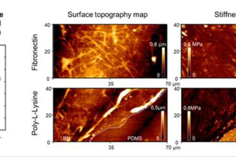
![Figure 5 from Catharina Husteden et al. 2023 “Lipoplex-Functionalized Thin-Film Surface Coating Based on Extracellular Matrix Components as Local Gene Delivery System to Control Osteogenic Stem Cell Differentiation”: A–D) Topography images [Cs/Col]4Cs (A), [Cs/Col]4Cs/LPX (B), [Cs/Col]4Cs/LPX/Cs (C), and [Cs/Col]4Cs/LPX/Cs/Col determined by AFM [Scale bar 1 µm] (D). E) Distribution curves of Young's modulus (E0) with a force map of a defined area (see also Figure S4, Supporting Information of the cited article). NANOSENSORSTM uniqprobe triangular qp-BioT AFM probes were used for the quantitative imaging to investigate the surface roughness and topography (in a standard liquid cell of a commercially available atomic force microscope) and force mapping. The uniqprobe qp-BioT AFM probe types offer an alternative to silicon nitride probes, with the advantage of taller AFM tips with smaller opening angles and reduced thermal drift.](https://d218f3btfcac6d.cloudfront.net/wp-content/uploads/2024/05/16205027/Fig-5-C-Husteden-et-al-2023-Lipoplex-Functionalized-Thin-Film-Surface-Coating-Based-on-Extracellular-Matrix-Components-NANOSENSORS-qp-BioT-AFM-probe-blog-930x620.jpg)

