Revisiting Contact Potential Difference in Electrostatic Force Microscopy
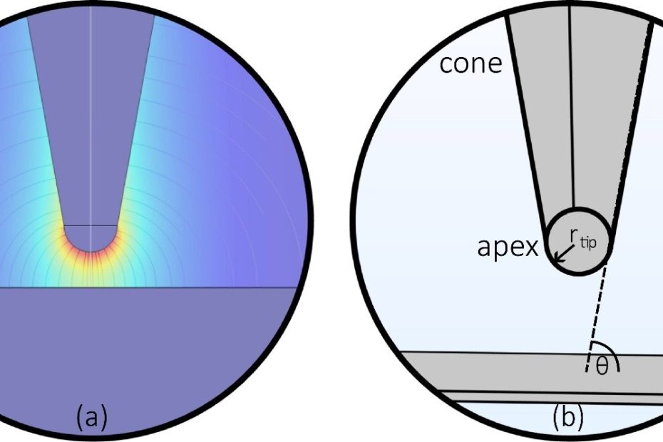
Electrostatic Force Microscopy (EFM) is an important atomic force microscopy (AFM) technique for investigating electrical properties and electrostatic interactions at the nanoscale. Achieving quantitative EFM… Read More »Revisiting Contact Potential Difference in Electrostatic Force Microscopy
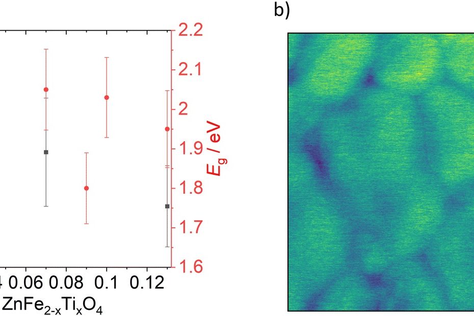


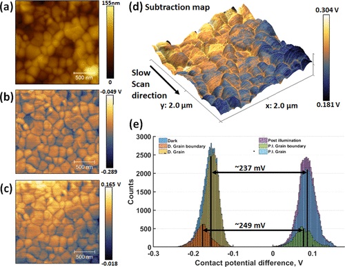
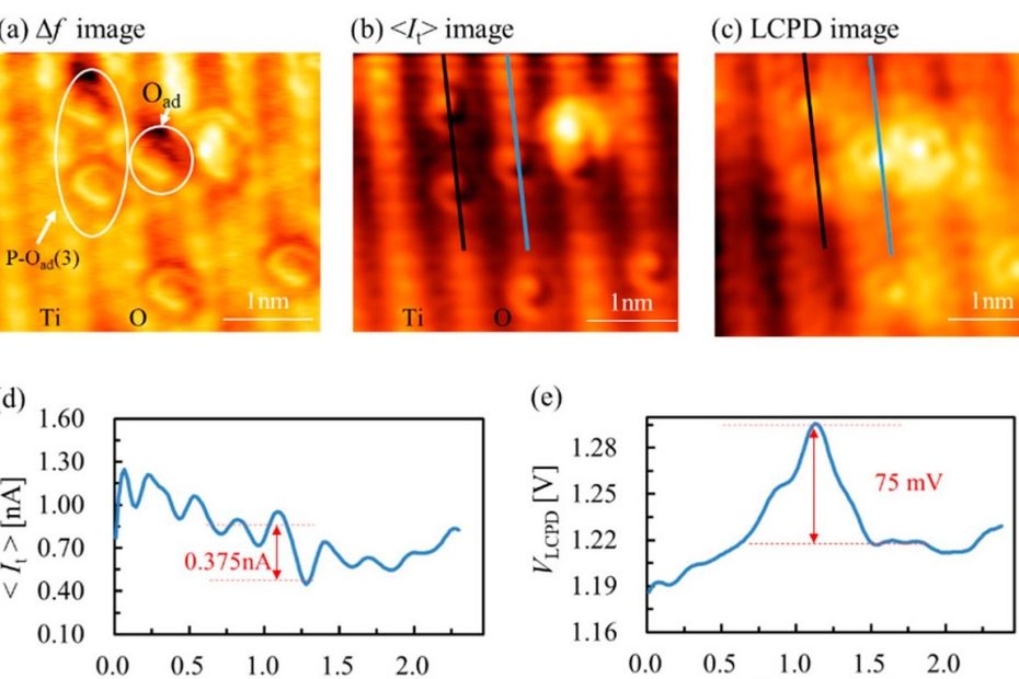
![Supplementary Figure 1 a – d from “Macroscopic manifestation of domain-wall magnetism and magnetoelectric effect in a Néel-type skyrmion host” by K. Geirhos et al: Typical ferroelectric do-main pattern observed on the (001) cleaved GaV4Se8 crystal surface atT=10 K. a, The topography is characterized by stripes roughly parallel to the [110] axis and folds parallel to the [010] axis. The latter originate in the differently oriented distortion of the ferroelastic domains. The color scale corresponds to the z-displacement of the tip. b ,In the dissipation channel of the nc-AFM every second domain appears bright. For the non-magnetic tip the dissipation originates from electric interactions. The dissipated power is indicated by the color scale. Please have a look at the full article to view the full supplementary figure. NANOSENSORS Platinum Silicide PtSi-FM AFM probes were used for the imaging.](https://www.nanosensors.com/blog/wp-content/uploads/2022/11/Supplementary-Figure-1-a-–-d-from-Macroscopic-manifestation-of-domain-wall-magnetism-and-magnetoelectric-effect-in-a-Neel-type-skyrmion-host-by-K-Geirhos-et-al-PtSi-FM-2-766x620.jpg)