In the article “Multi-Channel Exploration of O Adatom on TiO2(110) Surface by Scanning Probe Microscopy” Huan Fei Wen, Yasuhiro Sugawara and Yan Jun describe how they studied the O2 dissociated state under the different O2 exposed temperatures with atomic resolution by scanning probe microscopy (SPM) and imaged the O adatom by simultaneous atomic force microscopy (AFM)/scanning tunneling microscopy (STM).*
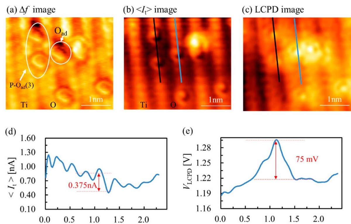
The effect of AFM operation mode on O adatom contrast was investigated, and the interaction of O adatom and the subsurface defect was observed by AFM/STM. Multi-channel exploration was performed to investigate the charge transfer between the adsorbed O and the TiO2(110) by obtaining the frequency shift, tunneling current and local contact potential difference at an atomic scale. The tunneling current image showed the difference of the tunneling possibility on the single O adatom and paired O adatoms, and the local contact potential difference distribution of the O-TiO2(110) surface institutively revealed the charge transfer from TiO2(110) surface to O adatom. The experimental results are expected to be helpful in investigating surface/interface properties by SPM. *
Iridium-coated ultrastiff AFM cantilever SD-T10L100 from the NANOSENSORS Special Developments List (typical Force constant 2 000 N/m) were used in the presented study.
The cantilever tip was first degassed at approximately 650 K for 30 min and then cleaned by Ar ion bombardment to remove the contaminants, prior to the measurements. Features of the surface structure were related to the charge states of the tip apex, and a stable tip was essential to accurately characterize the surface structure and properties in the experiment. The imaging mode became stable in AFM experiments when the metal-coated Si cantilever was employed in the experiments. *

Figure 5. from “Multiple images of TiO2(110) surface with atomic resolution and corresponding line profiles” by Huan Fei Wen et al.
(a) Frequency shift (∆f) image, (b) tunneling current (<It>) image and (c) local contact potential difference (VLCPD) image. (d,e) The line profiles along the blue line on the surface in (b,c). (f0 = 805 kHz, Q = 27623, ∆f = −260 Hz, VDC = 1.3 V, VAC = 1.5 V, A = 500 pm, size: 3.5 × 3.2 nm2). Multiple images of TiO2(110) surface with atomic resolution and corresponding line profiles. (a) Frequency shift (∆f) image, (b) tunneling current (<It>) image and (c) local contact potential difference (VLCPD) image. (d,e) The line profiles along the blue line on the surface in (b,c). (f0 = 805 kHz, Q = 27623, ∆f = −260 Hz, VDC = 1.3 V, VAC = 1.5 V, A = 500 pm, size: 3.5 × 3.2 nm2).
*Huan Fei Wen, Yasuhiro Sugawara and Yan Jun
Multi-Channel Exploration of O Adatom on TiO2(110) Surface by Scanning Probe Microscopy
Nanomaterials 2020, 10(8), 1506
DOI: https://doi.org/10.3390/nano10081506
Please follow this external link to read the full article: https://www.mdpi.com/2079-4991/10/8/1506/htm
More articles mentioning the use of NANOSENSORS ultrastiff AFM probes:
Yuuki Adachi, Huan Fei Wen, Quanzhen Zhang, Masato Miyazaki, Yasuhiro Sugawara and Yan Jun Li
Elucidating the charge state of an Au nanocluster on the oxidized/reduced rutile TiO2 (110) surface using non-contact atomic force microscopy and Kelvin probe force microscopy
Nanoscale Adv., 2020, 2, 2371-2375 (Paper)
DOI: 10.1039/C9NA00776H
https://pubs.rsc.org/en/content/articlehtml/2020/na/c9na00776h
Huan Fei Wen, Hongqian Sang Yasuhiro Sugawara, and Yan Jun Li
Dynamic behavior of OH and its atomic contrast with O adatom on the Ti site of TiO2(110) at 78 K by atomic force microscopy imaging
Appl. Phys. Lett. 117, 051602 (2020)
DOI: https://doi.org/10.1063/5.0016657
Yuuki Adachi, Yasuhiro Sugawara, and Yan Jun Li
Remotely Controlling the Charge State of Oxygen Adatoms on a Rutile TiO2(110) Surface Using Atomic Force Microscopy
J. Phys. Chem. C 2020, 124, 22, 12010–12015
DOI: https://doi.org/10.1021/acs.jpcc.0c03117
Huan Fei Wen, Quanzhen Zhang, Yuuki Adachi, Masato Miyazaki, Yasuhiro Sugawara, Yan JunLi
Contrast inversion of O adatom on rutile TiO2(1 1 0)-(1 × 1) surface by atomic force microscopy imaging
Applied Surface Science Volume 505, 1 March 2020, 144623
DOI: https://doi.org/10.1016/j.apsusc.2019.144623
Open Access The article “Multi-Channel Exploration of O Adatom on TiO2(110) Surface by Scanning Probe Microscopy” by Huan Fei Wen, Yasuhiro Sugawara and Yan Jun is licensed under a Creative Commons Attribution 4.0 International License, which permits use, sharing, adaptation, distribution and reproduction in any medium or format, as long as you give appropriate credit to the original author(s) and the source, provide a link to the Creative Commons license, and indicate if changes were made. The images or other third party material in this article are included in the article’s Creative Commons license, unless indicated otherwise in a credit line to the material. If material is not included in the article’s Creative Commons license and your intended use is not permitted by statutory regulation or exceeds the permitted use, you will need to obtain permission directly from the copyright holder. To view a copy of this license, visit http://creativecommons.org/licenses/by/4.0/.