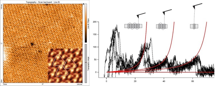
High resolution imaging of the cytoplasmic side of bacteriorhodospin (BR) (left) and overlay of 6 force-distance curves (right) showing the controlled C-terminal unfolding of a single BR membrane protein from its native environment.
The measurements were recorded in buffer solution with a FlexAFM V3 (Nanosurf) using a NANOSENSORS™ uniqprobe qp-CONT cantilever by Dr. Patrick Frederix from Nanosurf AG.
The left image shows a topography image of a 2D crystalline patch of BR (also called purple membrane). A cross-correlation average calculated from this topography image is shown in the inset. Three bacteriorhodopsin trimers are highlighted.
The overlay of multiple force-distance curves on the right picture shows the controlled C-terminal unfolding of single BR membrane proteins from their native environment. It is clearly visible that unfolding pathways vary from molecule to molecule, but main barriers exist, for instance where the structure enters the membrane.
NANOSENSORS™ uniqprobe qp-CONT probes are especially well adapted for such kind of measurements since they feature a sharp tip combined with the low force constant of the cantilever, a high deflection sensitivity and a reduced drift due to the partial gold coating.