Soluble fibrin (SF) in blood consists of monomers lacking both fibrinopeptides A with a minor population in multimeric clusters. It is a substantial component of isolated fibrinogen (fg), which spontaneously self-assembles into protofibrils progressing to fibers at sub-physiologic temperatures, a process enhanced by adsorption to hydrophobic and some metal surfaces. *
In their article “Novel characteristics of soluble fibrin: hypercoagulability and acceleration of blood sedimentation rate mediated by its generation of erythrocyte-linked fibers” Dennis K. Galanakis, Anna Protopopova, Kao Li, Yingjie Yu, Tahmeena Ahmed, Lisa Senzel, Ryan Heslin, Mohamed Gouda, Jaseung Koo, John Weisel, Marilyn Manco-Johnson and Miriam Rafailovich mention how they employed topographic and lateral atomic force microscopy (AFM) scanning for imaging fg monomers and soluble polymers. Adsorption to polystyrene (PS) and to trioctylmethylamine (TOMA) coated silica wafers was used.
NANOSENSORS™ SuperSharpSilicon™ SSS-SEIHR high resolution AFM probes (typical AFM tip radius of 2 nm, typical force constant 15 N/m, typical resonance frequency 130 kHz) were used for the atomic force microscopy (AFM) in tapping mode. *
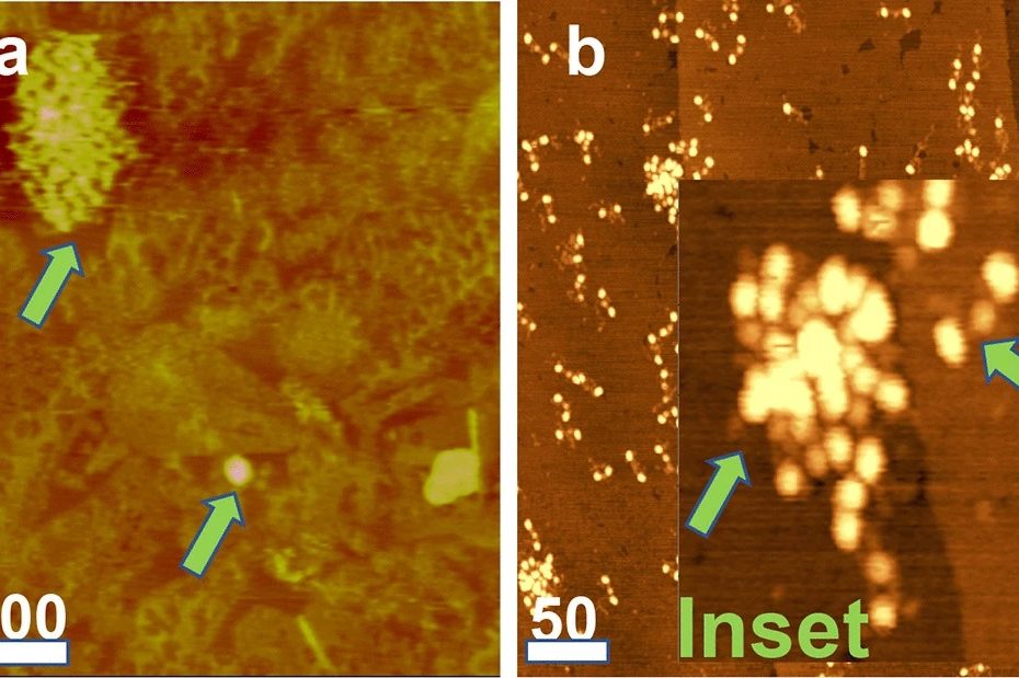
Figure 5 from D. Galanakis et al. “Novel characteristics of soluble fibrin: hypercoagulability and acceleration of blood sedimentation rate mediated by its generation of erythrocyte-linked fibers”: AFM images. Horizontal bars denote 100 nm and 50 nm in panels a and b, respectively. a FR fg adsorbed on a TOMA surface showing a large, upper arrow, and a smaller multimeric cluster, lower arrow, showing the marked variation in the cluster size. The smaller cluster also shows regular surface undulations indicating its multimeric composition. b FR fg adsorbed on modified graphite (MG) surface, showing a field of solitary trinocular monomers and two clusters. Inset: a 4 × magnification from a different area of the same field showing a monomer, right arrow, and a multimeric cluster, left arrow
*Dennis K. Galanakis, Anna Protopopova, Kao Li, Yingjie Yu, Tahmeena Ahmed, Lisa Senzel, Ryan Heslin, Mohamed Gouda, Jaseung Koo, John Weisel, Marilyn Manco-Johnson and Miriam Rafailovich
Novel characteristics of soluble fibrin: hypercoagulability and acceleration of blood sedimentation rate mediated by its generation of erythrocyte-linked fibers
Cell and Tissue Research 387, pages 479–491 (2022)
DOI: https://doi.org/10.1007/s00441-022-03599-9
Please follow this external link to read the full article: https://rdcu.be/cL9qn
Open Access: The article “Novel characteristics of soluble fibrin: hypercoagulability and acceleration of blood sedimentation rate mediated by its generation of erythrocyte-linked fibers” by Dennis K. Galanakis, Anna Protopopova, Kao Li, Yingjie Yu, Tahmeena Ahmed, Lisa Senzel, Ryan Heslin, Mohamed Gouda, Jaseung Koo, John Weisel, Marilyn Manco-Johnson and Miriam Rafailovich is licensed under a Creative Commons Attribution 4.0 International License, which permits use, sharing, adaptation, distribution and reproduction in any medium or format, as long as you give appropriate credit to the original author(s) and the source, provide a link to the Creative Commons license, and indicate if changes were made. The images or other third party material in this article are included in the article’s Creative Commons licence, unless indicated otherwise in a credit line to the material. If material is not included in the article’s Creative Commons licence and your intended use is not permitted by statutory regulation or exceeds the permitted use, you will need to obtain permission directly from the copyright holder. To view a copy of this licence, visit http://creativecommons.org/licenses/by/4.0/.