Muscle wasting is connected with changes in various cellular mechanisms that influence protein homeostasis, transcription, protein acetylation and different metabolic pathways. *
Scientific studies have shown that reduced levels of polyadenylation binding protein 1 ( PABPN1 , a multifactorial regulator of mRNA processing ) cause muscle wasting, including muscle atrophy, extracellular matrix thickening, myofiber typing transitions and central nucleation. *
However, the cellular mechanisms behind PABPN1-mediated muscle wasting are not fully understood. *
In the article “Cytoskeletal disorganization underlies PABPN1-mediated myogenic disability” Cyriel Sebastiaan Olie, Erik van der Wal, Cikes Domagoj, Loes Maton, Jessica C. de Greef, I.-Hsuan Lin, Yi-Fan Chen, Elsayad Kareem, Josef M. Penninger, Benedikt M. Kessler and Vered Raz examine the cytoskeletal auxiliary changes that are dependent on PABPN1 levels using 2D and 3D models, and investigate how these affect muscle wasting. *
They suggest that poor cytoskeletal mechanical features are caused by altered expression levels of cytoskeletal proteins and contribute to muscle wasting and atrophy. *
For the measurements of cell-mechanics properties in control and shPAB cells ( muscle cells with reduced PABPN1 levels ) the authors used Brillouin Light Scattering Microscopy and Atomic Force Microscopy. *
NANOSENSORS™ uniqprobe qp-BioAC ( CB3 ) AFM probes were used in the quantitative imaging where a force curve is applied at each point. The analyzed area of each cell was 5 µm × 5 µm (64 × 64 pixels) with an approach speed of 35 µm/s (3.4 ms/pixel), and applied forces of up to 118 pN. *
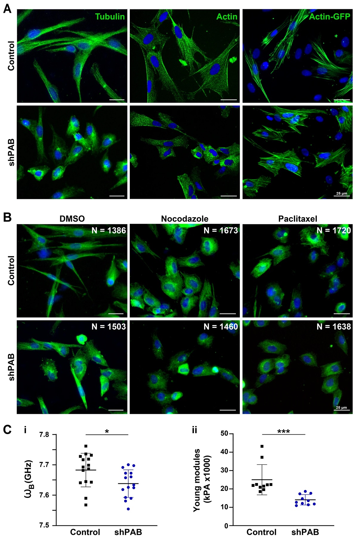
Disrupted cytoskeletal spatial organization in shPAB human muscle cell cultures. A Representative images of control and shPAB human muscle cell cultures, stained with antibodies to tubulin and actin, and the actin filaments were visualized with actin-GFP. B Tubulin staining in control and shPAB myoblast cell cultures after DMSO, 100 nM nocodazole or 25 nM paclitaxel treatment for 2 h. Scale bar is 25 µm. C Measurements of cell-mechanics properties in control and shPAB cells using the Brillouin Light Scattering Microscopy (Ci) or the Atomic Force Microscopy (Cii). Measurements were carried out in myoblasts; every dot represents the median from 1000 measurements in a cell. Cell stiffness is measured by GHz, and the young modulus reports the cell surface tension. Averages and standard deviations are from N = 15 cells. Statistical significance was calculated with the student’s t-test.
*Cyriel Sebastiaan Olie, Erik van der Wal, Cikes Domagoj, Loes Maton, Jessica C. de Greef, I.-Hsuan Lin, Yi-Fan Chen, Elsayad Kareem, Josef M. Penninger, Benedikt M. Kessler & Vered Raz
Cytoskeletal disorganization underlies PABPN1-mediated myogenic disability
Nature Scientific Reports volume 10, Article number: 17621 (2020)
DOI: https://doi.org/10.1038/s41598-020-74676-8
Please follow this external link to read the full article https://rdcu.be/ceyJ4
Open Access: The article “Cytoskeletal disorganization underlies PABPN1-mediated myogenic disability” by Cyriel Sebastiaan Olie, Erik van der Wal, Cikes Domagoj, Loes Maton, Jessica C. de Greef, I.-Hsuan Lin, Yi-Fan Chen, Elsayad Kareem, Josef M. Penninger, Benedikt M. Kessler & Vered Raz is licensed under a Creative Commons Attribution 4.0 International License, which permits use, sharing, adaptation, distribution and reproduction in any medium or format, as long as you give appropriate credit to the original author(s) and the source, provide a link to the Creative Commons license, and indicate if changes were made. The images or other third party material in this article are included in the article’s Creative Commons license, unless indicated otherwise in a credit line to the material. If material is not included in the article’s Creative Commons license and your intended use is not permitted by statutory regulation or exceeds the permitted use, you will need to obtain permission directly from the copyright holder. To view a copy of this license, visit http://creativecommons.org/licenses/by/4.0/.