SSS-FM

Cantilever data:
Property Nominal Value Specified Range
Resonance Frequency [kHz] 75 45 - 115
Force Constant [N/m] 2.8 0.5 - 9.5
Length [µm] 225 215 - 235
Mean Width [µm] 30 20 - 35
Thickness [µm] 3 2 - 4
Order codes and shipping units:
Order Code AFM probes per pack Data sheet
SSS-FM-10 10 of all probes
SSS-FM-20 20 of all probes
SSS-FM-50 50

Special handling information for NANOSENSORS™

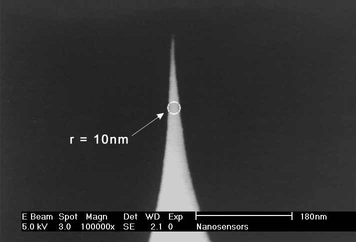
Due to their unique geometry the tips of the are more susceptible to tip damage by electrostatic discharge (ESD) than other Silicon-SPM-Probes.

Electric fields near the probe chip may lead to field evaporation which can blunt the tip apex of the probe tip. Therefore the NANOSENSORS™ are shipped in specially designed ESD-safe chip carriers.

NANOSENSORS™ recommends to their customers to take appropriate precautions to avoid tip damage due to electrostatic discharge when handling the probes. This can for example be done by using anti-electrostatic mats, wrist bands and tweezers.

SuperSharpSilicon™- Force Modulation Mode

NANOSENSORS™ SSS-FM AFM probes are designed for force modulation mode.

For enhanced resolution of nanostructures and microroughness we offer our SuperSharpSilicon™ AFM tip with unrivalled sharpness.

The FM type is offered for force modulation microscopy. The force constant of this AFM probe spans the gap between contact and non-contact mode and is specially tailored for the force modulation mode. The SSS-FM tip serves also as a basis for high resolution AFM tips with magnetic coatings (SSS-MFMR). Furthermore non-contact or tapping mode operation is possible with the FM tip but with reduced operation stability.

The AFM probe offers unique features:
  • guaranteed AFM tip radius of curvature < 5 nm
  • typical AFM tip radius of curvature of 2 nm
  • typical aspect ratio at 200 nm from tip apex in the order of 4:1
  • half cone angle at 200 nm from apex < 10°
  • monolithic material
  • highly doped to dissipate static charge
  • chemically inert
  • high mechanical Q-factor for high sensitivity

Szabo GL, Jany BR, Muckenhuber H, Niggas A, Lehner M, Janas A, Szabo PS, Gan Z, George A, Turchanin A, Krok F
Charge‐State‐Enhanced Ion Sputtering of Metallic Gold Nanoislands
Small. 2023 Jun;19(26):2207263
DOI: https://doi.org/10.1002/smll.202207263


Paul A, Rayabharam A, Murkute P, Almonte L, Rigo E, Dong Z, Kumar A, Joseph J, Aluru N, Timp G
Decoding Proteoforms with Single Acid Resolution Using a Sub-nanometer Diameter Pore
bioRxiv. 2022 Dec 22:2022-12
DOI: https://doi.org/10.1101/2022.12.22.521660


Sarveswaran K, Kurz V, Dong Z, Tanaka T, Penny S, Timp G
Synthetic capillaries to control microscopic blood flow
Scientific reports. 2016 Feb 24;6(1):21885
DOI: https://doi.org/10.1038/srep21885


Harte NP, Klyubin I, McCarthy EK, Min S, Garrahy SA, Xie Y, Davey GP, Boland JJ, Rowan MJ, Mok KH
Amyloid oligomers and mature fibrils prepared from an innocuous protein cause diverging cellular death mechanisms.
Journal of Biological Chemistry. 2015 Nov 20;290(47):28343-52
DOI: https://doi.org/10.1074/jbc.M115.676072


Mücke N, Kirmse R, Wedig T, Leterrier JF, Kreplak L.
Investigation of the morphology of intermediate filaments adsorbed to different solid supports
Journal of structural biology. 2005 Jun 1;150(3):268-76
DOI: https://doi.org/10.1016/j.jsb.2005.02.012