AR5-NCLR

Cantilever data:
Property Nominal Value Specified Range
Resonance Frequency [kHz] 190 146 - 236
Force Constant [N/m] 48 21 - 98
Length [µm] 225 215 - 235
Mean Width [µm] 38 30 - 45
Thickness [µm] 7 6 - 8
Order codes and shipping units:
Order Code AFM probes per pack Data sheet
AR5-NCLR-10 10 of all probes
AR5-NCLR-20 20 of all probes
AR5-NCLR-50 50
AR5-NCLR-W 370

Special handling information for NANOSENSORS™

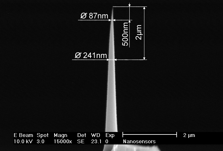
Due to their unique geometry the tips of the are more susceptible to tip damage by electrostatic discharge (ESD) than other Silicon-SPM-Probes.

Electric fields near the probe chip may lead to field evaporation which can blunt the tip apex of the probe tip. Therefore the NANOSENSORS™ are shipped in specially designed ESD-safe chip carriers.

NANOSENSORS™ recommends to their customers to take appropriate precautions to avoid tip damage due to electrostatic discharge when handling the probes. This can for example be done by using anti-electrostatic mats, wrist bands and tweezers.

NANOSENSORS™ High Aspect Ratio Silicon AFM probes

High Aspect Ratio (> 5:1) - Non-Contact/Tapping Mode - Long Cantilever - Reflex Coating

NANOSENSORS™ AR5-NCLR AFM tips are designed for non-contact or tapping mode AFM. The NCL probe is designed for SPM systems requiring a minimum AFM cantilever length > 125 µm or a resonance frequency of less than 400 kHz. Compared to the high frequency non-contact type NCH the maximum scanning speed is slightly reduced. This AFM cantilever type combines high operation stability with outstanding sensitivity and fast scanning ability.

For measurements on samples with sidewall angles approaching 90° NANOSENSORS™ produces specially tailored AFM tips. These AFM tips are FIB (Focused Ion Beam) milled to achieve a high aspect ratio portion better than 5:1 at the end of the common silicon AFM tip. This subtractive method of producing the high aspect ratio needle offers the advantage of high lateral stiffness and rigidity of the AFM tip.

The probe offers unique features:

  • length of the high aspect ratio portion of the AFM tip > 2 µm
  • typical aspect ratio at 2 µm in the order of 7:1 (when viewed from side as well as along AFM cantilever axis)
  • half cone angle at 2 µm of the high aspect ratio portion typically < 5°
  • guaranteed AFM tip radius of curvature < 15 nm
  • highly doped silicon to dissipate static charge
  • high mechanical Q-factor for high sensitivity
  • alignment grooves on backside of silicon holder chip
  • precise alignment of the AFM cantilever position (within +/- 2 µm) when used with the Alignment Chip
  • compatible with PointProbe® Plus XY-Alignment Series
This AFM probe features alignment grooves on the back side of the holder chip. These grooves fit to the NANOSENSORS Alignment Chip.

Xin CJ, Lu S, Yang J, Shams-Ansari A, Desiatov B, Magalhães LS, Ghosh SS, McGee E, Renaud D, Achuthan N, Zvyagintsev A
Wavelength-accurate and wafer-scale process for nonlinear frequency mixers in thin-film lithium niobate
Communications Physics. 2025 Apr 6;8(1):136
DOI: https://doi.org/10.1038/s42005-025-02068-3


Mukaddam K, Astasov-Frauenhoffer M, Fasler-Kan E, Marot L, Kisiel M, Steiner R, Sanchez F, Meyer E, Köser J, Bornstein MM, Kühl S
Novel titanium nanospike structure using low-energy Helium ion bombardment for the transgingival part of a dental implant
Nanomaterials. 2022 Mar 24;12(7):1065
DOI: https://doi.org/10.3390/nano12071065


Alsabti NI, Bourauel CP, Talic NF
Comparison of surface topography of low-friction and conventional TMA orthodontic arch wires using atomic force microscopy
Journal of Orthodontic Science. 2021 Jan 1;10(1):2
DOI: https://doi.org/10.4103/jos.JOS_27_20


Singh M, Guzman-Aranguez A, Hussain A, Srinivas CS, Kaur IP
Solid lipid nanoparticles for ocular delivery of isoniazid: evaluation, proof of concept and in vivo safety & kinetics
Nanomedicine. 2019 Feb 1;14(4):465-91
DOI: https://doi.org/10.2217/nnm-2018-0278


Azeem A, English A, Kumar P, Satyam A, Biggs M, Jones E, Tripathi B, Basu N, Henkel J, Vaquette C, Rooney N
The influence of anisotropic nano-to micro-topography on in vitro and in vivo osteogenesis
Nanomedicine. 2015 Mar 1;10(5):693-711
DOI: https://doi.org/10.2217/nnm.14.218